
No plaster cast
Your baby's head is never cast in plaster. Measurement is completed in a single session with a contactless 3D scan that takes seconds.
The treatment window is short, so waiting time matters.
The optimal window for treating plagiocephaly and brachycephaly is months 4 to 8. With Clonify, a contactless scan replaces the plaster mould; measurement and helmet design happen in the app, and the production STL is released after clinician approval. The wait is shorter because the moulding and manual shaping steps are gone.
Helmet therapy works with the infant's rapid skull growth. The optimal window is months 4–8, and every week spent in the plaster process comes out of it. Outcomes are markedly better when treatment starts early.
Months 4–8
Optimal treatment window
10–18 days
Average delivery in the plaster process
No plaster
Contactless scan with Clonify, one session
For families
Flat head syndrome (plagiocephaly) is common in infants and can be managed successfully when addressed at the right time. In clinics using Clonify, the process is far more comfortable for your baby.
Your baby's head is never cast in plaster. Measurement is completed in a single session with a contactless 3D scan that takes seconds.
Head asymmetry is measured millimetrically with international metrics such as CVAI and cephalic index — and reported to you.
At every check-up, the new scan is automatically compared with the previous one; you follow your baby's progress in numbers and visuals.
Because the treatment window is narrow, speed matters. The digital process targets helmet delivery within days.
The four views below map to desktop workspace steps. The numbers are from the sample report; the mesh is a downsampled PLY of the same case.
Drag to rotate the model · Stages advance as you scroll
01 · Import
The head scan loads as PLY or STL. The point cloud on screen is made of the sample file's real vertices.
Real sample scan — measurements from the 19 Aug 2026 cranial report
The head scan loads as PLY or STL. The point cloud on screen is made of the sample file's real vertices.
After alignment, CVAI, cephalic index, CVA and circumference are measured on Z slices. The heat map compares the two sides of the mid-sagittal plane and shows flattened and bossed areas.
A future head is built from target CVAI/CI. To leave room for growth, the helmet's inner surface stays at least 10 mm outside the scan everywhere.
In this step the fit is checked and the orthotist approves; the clinical PDF and STL are then exported. Zones (1.5–2.5 mm active / 6–8 mm passive) and assembly (holes, lid, hinge) finish the helmet.
Head Socket Analyses
The scan is imported, aligned to the anatomical plane, sliced, photographed and compared with a previous scan. The PDF report is also produced here.
Import
An open-format (PLY/STL/OBJ/CPX/VTP) head scan is loaded; smoothing, densification and hole/shell repair run in this step.
Head Alignment
The model is aligned to the anatomical plane. Ear landmarks are the reference for later trim and helmet lines.
Head Slicer
CVAI, cephalic index, circumference and diameters are measured on Z slices. Sample report: CVAI 6.1% · CI 87.6% · CVA 8.6 mm · circ. 438.1 mm.
Cranial Photo Capture
Clinical photographs are bound to the same file as the scan and embedded in the report.
Head Comparison
Two scans side by side: anthropometry, asymmetry, volume and a live slice. The PDF report is produced here.
Head Socket Design
From the aligned scan: target shape, clearance, shell, assembly and STL export. CVAI/CI are measured during helmet design.
Import
The same scan file is opened for helmet design.
Head Alignment
Alignment from analysis is kept or repeated.
Cranial
The current head is measured. A future head is built from target CVAI/CI; the helmet inner surface stays at least 10 mm outside the scan (growth allowance).
Zone / Clearance
Active (1.5–2.5 mm) and passive (6–8 mm) clearance bands are applied to the target shape.
Shell / Offset
The liner inner-surface offset and an editable trim loop are set; the cut itself is made separately with Apply Crop.
Assembly
Tools for holes, the lid, a dual symmetric lock mechanism and a circumferential separation cut sit in this step, each on its own tab.
Validate / Export
Fit check, orthotist approval, clinical report and STL export.
For clinicians
Many clinics use separate programs for analysis and design. In Clonify, measurement is automatic, the clinician approves the helmet design, and the report for the family or physician comes from the same case.
The head model aligns automatically to the anatomical plane, and CVAI, cephalic index, circumference and diameters are calculated automatically. Severity classification and clinical decisions belong to the clinician.
Pre- and post-treatment scans are compared automatically. A PDF or Excel report takes one click, and you can share it with the referring physician.
The software proposes the design step by step. You decide on relief and pressure zones and the trimline.
Our target: ≤8 hours of training, first unassisted device within ≤5 workdays. We co-design the first 10 devices with you remotely.
Comparison
Plaster workshop on the left, digital workflow on the right.
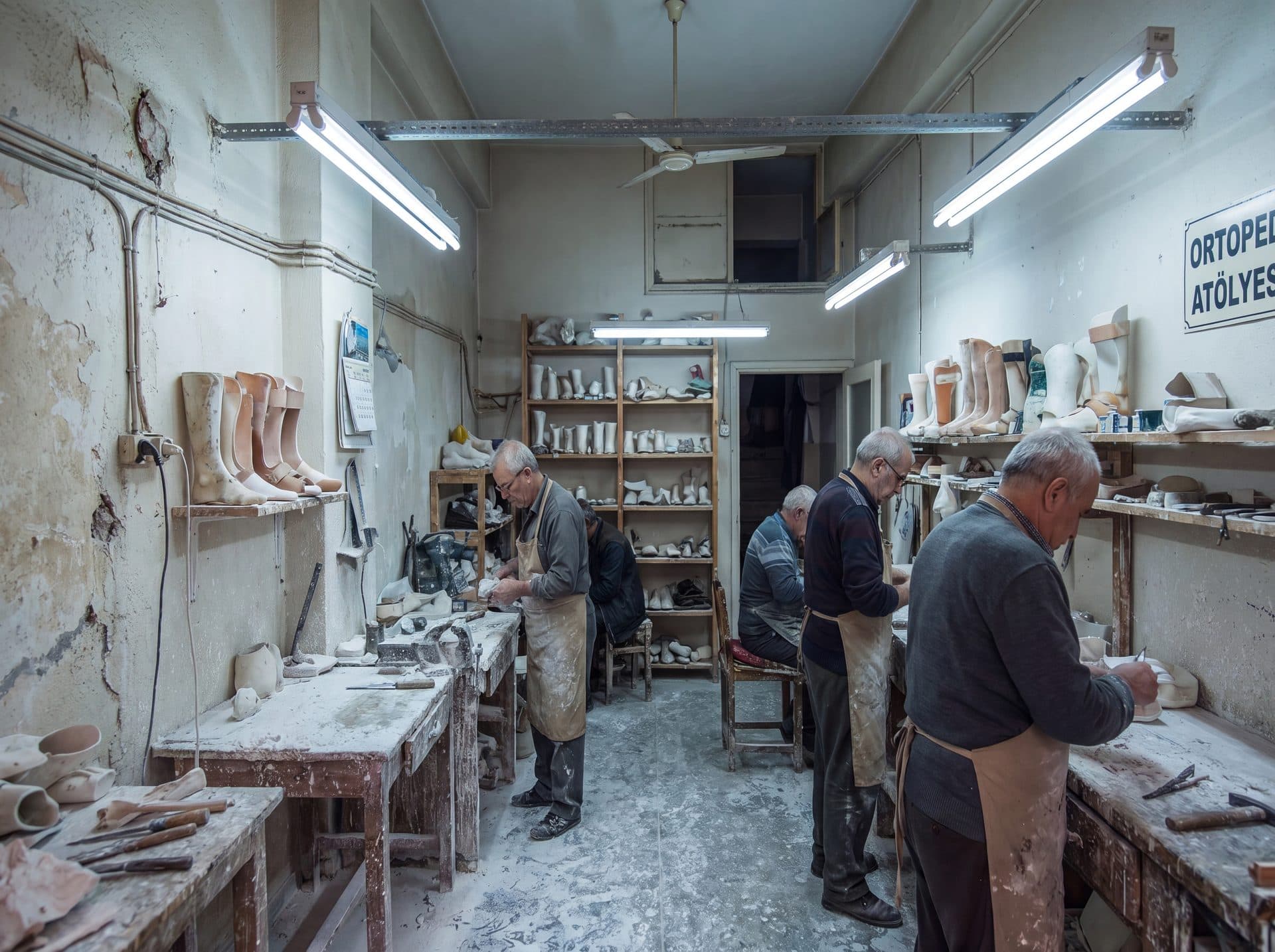
Plaster process
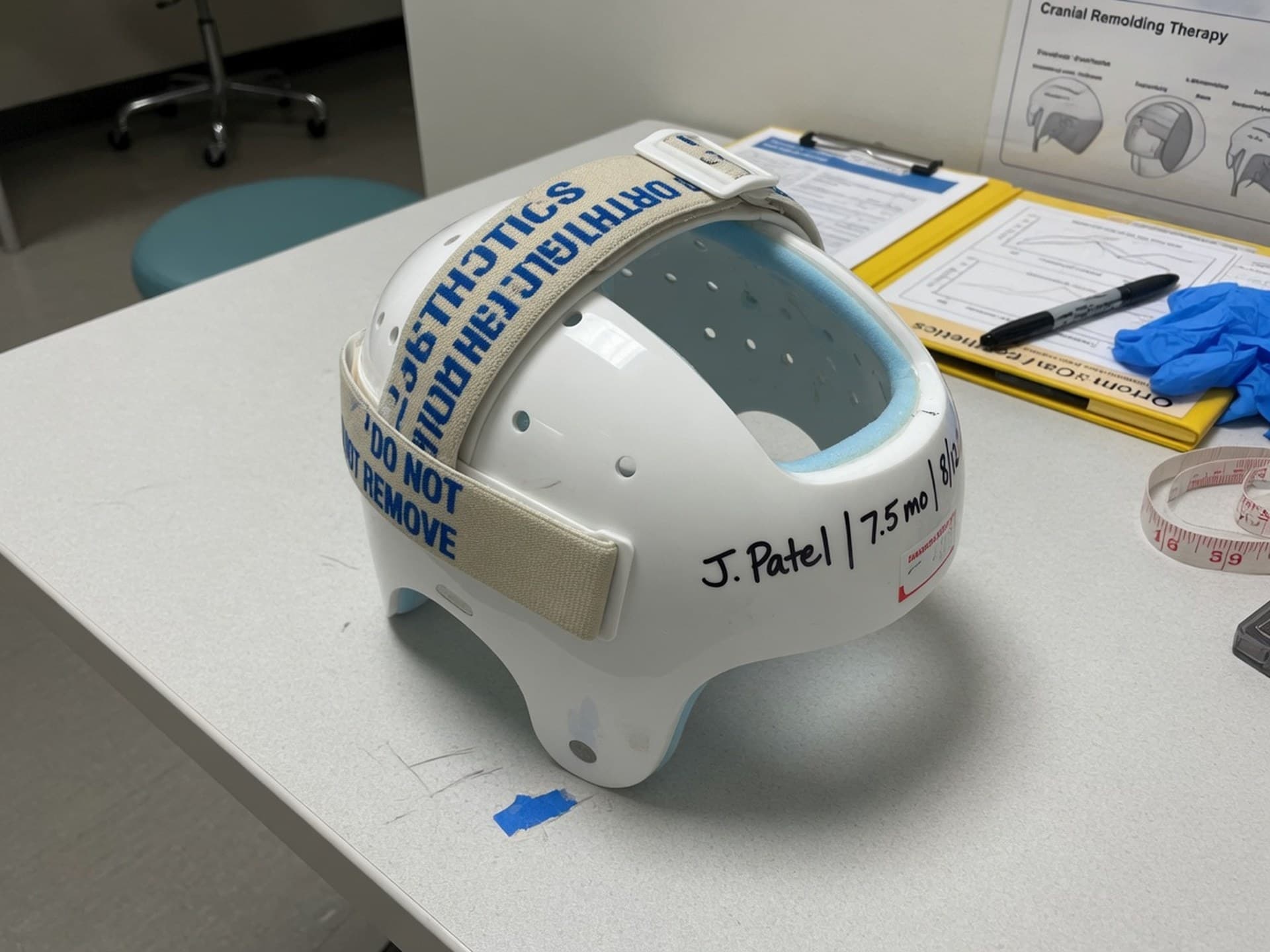
Clonify process
Sample clinical report
Clonify desktop export dated 19 Aug 2026 (kafa1.ply). The name line is the sample 'example'; this is not a diagnosis. Measured values: CVAI 6.1% · cephalic index 87.6% · CVA 8.6 mm · slice circumference 438.1 mm. Severity classification is the clinician's call.
| Metric | Value | Note |
|---|---|---|
| CVAI | 6.1% | Measured value — classification is clinical |
| Cephalic index | 87.6% | ML 129.1 / AP 147.4 mm |
| CVA | 8.6 mm | A 149.8 · B 141.2 |
| Circumference | 438.1 mm | AP 147.4 · ML 129.1 |
The report is exported as PDF from the desktop app and shared with the physician and family.
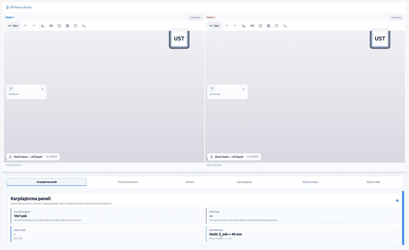
Bring a scan of your own patient to the demo; see the analysis report and design flow on your own case.
The plaster-era delivery time is a field average. Delivery time with Clonify is being measured in pilot clinics; results will be published on the Evidence page. The treatment decision always belongs to your physician and clinician.
Sockets, splints and the full workflow: O&P CAD software guide
Sits on the scanner and printer you already have
Production files only after clinician approval
Built for custom-made device regulation